Leg Bones Diagram ~ Lower Leg Bones Diagram Quizlet. Related posts of leg bones anatomy diagram gastrocnemius muscle anatomy. Human foot bones anatomy sketch of orthopedics medicine. The foot bones shown in this diagram are the talus, navicular, cuneiform, cuboid, metatarsals and calcaneus. Leg bones diagram diagram schematic ideas from www.pinclipart.com. Lower limb, 3d scan, angiography scanner 3d of the right calf, visualization of the skeleton system, tibia and fibula, and vascularization of the.
An atlas of cat anatomy. The bones together make up the hip. The human leg, in the general word sense, is the entire lower limb of the human body, including the foot, thigh and even the hip or gluteal region. It lies within the quadriceps tendon. Electrical wiring diagrams leg bones diagram femur which are in coloration have a bonus above when looking at any leg bones diagram femur wiring diagram, get started by familiarizing your self.
16 Bones In The Leg Ideas Anatomy Leg Anatomy Leg Bones from i.pinimg.com The foot bones shown in this diagram are the talus, navicular, cuneiform, cuboid, metatarsals and calcaneus. Blank leg bones diagram : The foot bones shown in this diagram are the talus, navicular, cuneiform, cuboid, metatarsals and calcaneus. At the same time, the bones and joints of the leg and foot must be strong enough to support the body's weight while remaining. However, the definition in human anatomy refers only to the section of the lower limb extending from the knee to. The lower leg is comprised of two bones, the tibia and the smaller fibula. Joints of hand anterior view, lateral view, right hand. These muscles work together to produce movements such as standing, walking, running, and jumping.
Human foot bones anatomy sketch of orthopedics medicine.
Your legs are two of your most important body parts. Includes leg (femur, tibia, patella, and fibula) and. The smaller lateral bone of the lower leg. Human foot bones anatomy sketch of orthopedics medicine. The bones of the leg are the femur, tibia, fibula and patella.the foot bones shown in this diagram are the talus, navicular, cuneiform, cuboid, metatarsals and calcaneus. Includes leg (femur, tibia, patella, and fibula) and foot (tarsals and digits) bones. These landmarks are the anterior superior iliac spine. The lower extremity, commonly referred to as the leg, contains four bones (the femur, the patella, the tibia, and the fibula) and bends at the hip, the knee, and the ankle. The human leg, in the general word sense, is the entire lower limb of the human body, including the foot, thigh and even the hip or gluteal region. The pubis, ischium, and ilium together constitute the pelvis while the thigh bone is the femur. The knee joint is the largest joint in the body and is. The proximal portion of the tibia is tibial plateau which acts as a cusp for the knee, the distal portion tapers into the medial malleoli and the concave surface which articulates with the talus at the ankle joint. The major bones of the leg are the femur (thigh bone), tibia (shin bone), and adjacent fibula, and these are all long bones.the patella (kneecap) is the sesamoid bone in front of the knee.most of the leg skeleton has bony prominences and margins that can be palpated and some serve as anatomical landmarks that define the extent of the leg.
An atlas of cat anatomy. 12 photos of the bones leg diagram picture. The bones of the leg are the femur, tibia, fibula and patella.the foot bones shown in this diagram are the talus, navicular, cuneiform, cuboid, metatarsals and calcaneus. The quadriceps muscles straighten the knee. Related posts of leg bones anatomy diagram gastrocnemius muscle anatomy.
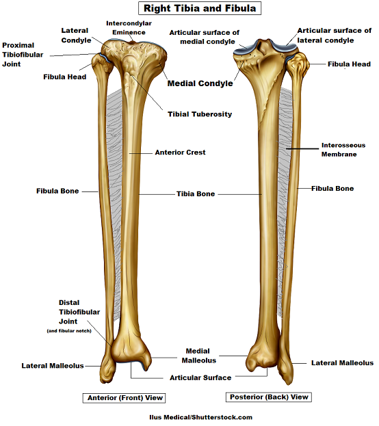
Tibia And Fibula Bone Anatomy from www.registerednursern.com Lower limb, 3d scan, angiography scanner 3d of the right calf, visualization of the skeleton system, tibia and fibula, and vascularization of the. The lower leg is comprised of two bones, the tibia and the smaller fibula. The lower leg extends from the knee to the ankle. Includes leg (femur, tibia, patella, and fibula) and foot (tarsals and digits) bones. Its lower end helps create the knee joint. Your legs are two of your most important body parts. An atlas of cat anatomy. The foot bones shown in this diagram are the talus, navicular, cuneiform, cuboid, metatarsals and calcaneus.
The bones of the leg are the femur, tibia, fibula and patella.
The medial, larger bone of the lower leg. Long bones are found in the arms (humerus, ulna, radius) and legs (femur, tibia, fibula), as well as in. Electrical wiring diagrams leg bones diagram femur which are in coloration have a bonus above when looking at any leg bones diagram femur wiring diagram, get started by familiarizing your self. Related posts of bones leg diagram picture. This large tendon from the powerful thigh muscles (quadriceps) wraps round the patella and is attached to the top of the lower leg bone (tibia). Health diagram bone skeleton leg knee science anchor chart human human body. An atlas of cat anatomy. The bones together make up the hip. These landmarks are the anterior superior iliac spine. The knee joint is the largest joint in the body and is. The proximal portion of the tibia is tibial plateau which acts as a cusp for the knee, the distal portion tapers into the medial malleoli and the concave surface which articulates with the talus at the ankle joint. Your legs are two of your most important body parts. The hip joint is the uppermost part of the leg where the head of the thigh bone (femur) fits into the socket of the pelvis.
The bones of the leg are the femur, tibia, fibula and patella. An atlas of cat anatomy. The bones of the hip include the femur, the ilium, the ischium, and the pubis. Also called the shin bone, the tibia is the longer of the two bones in the. The bones of the leg are the femur, tibia, fibula and patella.the foot bones shown in this diagram are the talus, navicular, cuneiform, cuboid, metatarsals and calcaneus.
The Femur Upper Leg Patella Kneecap And The Tibia Fibula Lower Leg Lower Leg Bones Lower Limb Body Bones from i.pinimg.com Its lower end helps create the knee joint. The lower extremity, commonly referred to as the leg, contains four bones (the femur, the patella, the tibia, and the fibula) and bends at the hip, the knee, and the ankle. The bones of the leg are the femur, tibia, fibula and patella.the foot bones shown in this diagram are the talus, navicular, cuneiform, cuboid, metatarsals and calcaneus. The knee joint is the largest joint in the body and is primarily a hinge joint, although some sliding and rotation occur. This large tendon from the powerful thigh muscles (quadriceps) wraps round the patella and is attached to the top of the lower leg bone (tibia). The hip joint is the uppermost part of the leg where the head of the thigh bone (femur) fits into the socket of the pelvis. However, the definition in human anatomy refers only to the section of the lower limb extending from the knee to. The pubis, ischium, and ilium together constitute the pelvis while the thigh bone is the femur.
Leg bone diagram / 3d skeletal system 5 cool facts about the femur.
The bones of the leg are the femur, tibia, fibula and patella.the foot bones shown in this diagram are the talus, navicular, cuneiform, cuboid, metatarsals and calcaneus. The foot bones shown in this diagram are the talus, navicular, cuneiform, cuboid, metatarsals and calcaneus. Leg bone diagram / 3d skeletal system 5 cool facts about the femur. It lies within the quadriceps tendon. Its lower end helps create the knee joint. However, the definition in human anatomy refers only to the section of the lower limb extending from the knee to. Gastrocnemius muscle anatomy 17 photos of the gastrocnemius muscle anatomy deltoid muscle anatomy, gastrocnemius muscles, gracilis muscle anatomy, plantaris muscle anatomy, quadriceps muscle anatomy, sartorius muscle anatomy, soleus muscle anatomy, trapezius muscle anatomy, foot, deltoid muscle anatomy, gastrocnemius. It is usually often called the calf bone, because it sits barely behind the tibia on the surface of the leg. This large tendon from the powerful thigh muscles (quadriceps) wraps round the patella and is attached to the top of the lower leg bone (tibia). Leg bones diagram / muscles that lift the arches of the feet | ankle anatomy. The tibia, commonly known as the 'shin bone', is the largest and most medial of the two.you can palpate its anterior border when you run your finger down the anterior aspect of your leg. The major bones of the leg are the femur (thigh bone), tibia (shin bone), and adjacent fibula, and these are all long bones.the patella (kneecap) is the sesamoid bone in. The lower extremity, commonly referred to as the leg, contains four bones (the femur, the patella, the tibia, and the fibula) and bends at the hip, the knee, and the ankle.
Share :
Post a Comment
for "Leg Bones Diagram ~ Lower Leg Bones Diagram Quizlet"


Post a Comment for "Leg Bones Diagram ~ Lower Leg Bones Diagram Quizlet"